Photos by Angela Shoemaker
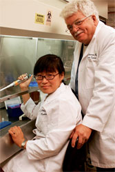
Dr. Ben Jenson and Shin-Je Ghim
 Ask Dr. Ben Jenson of the James Graham Brown Cancer Center how it feels to be on the team that discovered the first effective cancer vaccine and he’ll say, "It is difficult to describe completely. It’s like 15 years of butting one’s head against a wall, slogging through a bog, and finally reaching the /files/storyimages/of the tunnel where the light is."
Jenson, 67, and his U of L colleague, Shin-Je Ghim, 47, who has a doctorate in microbiology, believe the discovery — based on groundbreaking research they began in 1989 as part of a group at their former institution, Georgetown University — can render ineffective the human papillomavirus (HPV), strains of which cause most cases of cervical cancer. This virus is the most prevalent sexually transmitted disease worldwide, and cervical cancer is the world’s second-leading fatal cancer in women. (The U.S. mortality rate is much lower than in undeveloped countries because women here are better-screened for cervical abnormalities.)
In June, the FDA approved use of the vaccine for girls and women ages 9 to 26. Merck & Co. manufactures it under the name Gardasil. And while Georgetown holds the main patent on the drug, U of L’s research duo shares mightily in the result.
Through genetically engineered proteins, the Gardasil vaccine fools the body into thinking it is the virus. The body then unleashes an antibody response that neutralizes the real virus, Jenson says. Testing and FDA clinical trials show the vaccine is 100 percent effective in preventing cervical cancer, he says.
But there’s a problem: A three-dose vaccination costs approximately $350. In an effort to reduce that price, Jenson and Ghim are now focusing on an innovative source for a similar vaccine: tobacco leaves. In the next five years, Ghim says, she and Jenson hope to develop a genetically engineered tobacco "virus" that can be used to create a comparable, much less expensive vaccine. "Tobacco, which has carcinogenic properties, could also be used as part of a life-saving vaccine," Jenson says.
Meanwhile, the Centers for Disease Control and Prevention are expected to soon make recommendations on Gardasil, possibly paving the way for it to be prescribed as a medical option for young women. "I’m excited, because our work will help so many people," Ghim says. If it’s approved by the CDC, insurance companies would help pay for the cost of the vaccine, making it available to more women.
Dr. Lindsey Henson
 The "patient" is suffering from cardiac arrest, and University of Louisville medical students are performing resuscitation. Risky, you might say — until you learn that this patient is actually a mannequin programmed to respond like a human. Dr. Lindsey Henson, 57, interim director of the Dr. John M. and Dorothy S. Paris Simulation Center at U of L, says the med school’s surrogate sufferers allow students and staff to learn proper procedures in a risk-free environment.
While other medical schools and hospitals around the country train on these devices now, U of L was a trailblazer in this arena five years ago and still boasts one of the largest facilities in North America. In fact, the lab was featured in a recent segment on the Discovery Channel. It includes four simulation labs equipped with four programmable human-patient simulators and accompanying audio-visual capabilities.
During their first three years of training, all medical students are required to use the center on a regular basis. Dental and nursing students are also taught on the mannequins. For Henson, also chairman of the Department of Anesthesiology, one of most important roles of the devices is for those training in her specialty, who must practice on the high-tech mannequins for one intensive week before being allowed to work with living patients.
"One of the most important practical benefits of using the simulated patients is that students can work on their clinical skills in a risk-free environment, which gives them both technical skills and additional confidence when they begin working with patients," Henson says.
The human-patient simulators can be programmed with a multitude of medical problems. Students treat the "patient" while their instructors observe through a two-way mirror. If something goes wrong — or if the "patient" dies during the treatment — feedback is given as to what happened and why.
Henson says this practical approach to teaching and learning has earned U of L a reputation for excellence in the clinical area. "We feel it’s had a big impact on the medical education of our students,"
she says.
Within a decade, according to Henson, simulation training could well be required as a qualification for a medical license — much as airline pilots now practice on simulators before being allowed into the open sky.
Drs. Suzanne Ildstad and Warren Breidenbach
 Seeking to speed up advances in soft-tissue transplantation (skin, muscles, tendons, etc.) to benefit U.S. soldiers with burn/blast injuries, the Department of Defense recently awarded Jewish Hospital a $3.5 million grant (on top of a $2.5 million allocation in 2004) for hand and microsurgery research. Heading up the team of researchers, surgeons and support personnel involved in the project are Dr. Suzanne Ildstad, a transplant surgeon who also directs the University of Louisville’s Institute for Cellular Therapeutics, and Dr. Warren Breidenbach, the pioneering Kleinert Kutz surgeon who in 1999 performed the world’s second and third successful hand transplants.
Because any organ or tissue transplant requires recipients to ingest a multitude of anti-rejection immunosuppressive drugs daily for the rest of their lives — drugs that can cost $15,000-$25,000 a year, cause osteoporosis and diabetes, and weaken the body’s ability to fight off infections — the undertaking’s long-term goal is to eliminate the need for immunosuppression. How? By using bone-marrow stem cells retrieved from the donor to "trick" the host’s body into accepting the donor’s cells as its own. A first step — minimizing anti-rejection drugs through limited stem-cell use — is under way.
So far, Ildstad says, the marrow-replacement procedure has been successful in treating leukemia and, to a lesser degree, sickle-cell disease, and she’s optimistic about its current gradual implementation in heart and kidney transplants. "We don’t quite have our home run in the kidneys and hearts," she says, "but we’re seeing very promising results that suggest we’re very close."
Once the blood-disease and organ-transplantation phases are complete, the Ildstad/Breidenbach team will be able to take what they’ve learned and apply it to hand and other soft-tissue transplantation.
Dr. Paul Rychwalski
 When pediatric patients are brought to Kosair Children’s Hospital with suspected signs of abuse, a high-resolution digital camera known as a RetCam now offers physicians such as Dr. Paul Rychwalski the opportunity to document injuries in a new and more powerful way. Rychwalski, 39, and his colleagues can spot Shaken Baby Syndrome, which often results in traumatic eye injuries, including permanent forms of vision loss in 70 percent of victims. Astoundingly, in some cases, infants or children appear to be unaffected at first, but are later found to have signs of retinal hemorrhages, which can be spotted by the RetCam.
The new tool, purchased in 2002, allows larger areas of the retina and optic nerve to be photographed in 30-degree, wide-angle slices. These photographs can be shared, via computer, with other experts anywhere in the country, replacing the hand-drawn images of eye damage physicians have been using to depict Shaken Baby Syndrome, making them, among other uses, much more compelling evidence in a courtroom. Rychwalski, the hospital’s chief of pediatric ophthalmology and an assistant professor at U of L, sometimes testifies in court, and he says the images are harder to refute during trials of alleged child-abusers.
In addition to abuse injuries, the camera can detect abnormalities of children’s eyes. Retinoblastoma, a cancer, can be charted and tumor sizes from this condition monitored for better treatment. A congenital disorder called "retinopathy of prematurity" can be documented as well. In this condition, an infant’s premature delivery hinders the development of the retina, causing vision problems.
While the RetCam has numerous benefits to patients and physicians, there are limitations, Rychwalski says. One is that it requires contact with the eye’s surface, so in certain cases children might need sedation to allow photography. Another is cost: Kosair is now planning the purchase of another RetCam, this one smaller and more easily portable, but the price tag will be about $75,000, approximately $30,000 more expensive than the current instrument. But Rychwalski and his colleagues are excited about the instrument’s possibilities. They’re hoping to help bring the RetCam to Romania, where eye disorders are epidemic.
Dr. J. Michael Benfield
 On a typical day, a typical physician might see 25 patients in his office and sp/files/storyimages/15 minutes with each one. Dr. J. Michael Benfield is not a typical physician.
The founder of MD2U makes house calls to about 12 patients per day at their private residences and assisted-living addresses. On the initial visit, he spends between 60 and 90 minutes with a patient, with 30 minute follow-up visits at four-to-five-week intervals.
MD2U (on the internet as md2u.net) was born when Benfield, 40, sought a way to participate in nontraditional health care in a very "traditional" way — by helping revive the notion of doctor visits. "Home health nursing knew about the tremendous need of home-bound patients long ago," he says. A University of North Carolina graduate with a background in sales and marketing, he furthered his training at the East Carolina School of Medicine in 2000 and completed his residency at the University of Louisville in 2004. MD2U was launched shortly thereafter.
Benfield is currently the sole physician on staff; a nurse practitioner will be added this summer, he says. To keep costs low, the doctor’s wife and other family members help run the office, which is operated out of his home. Nearly all of his current patients are age 70 and above, and Benfield says he can provide them with a high level of care, in part by using clues from their home environments. "I can see what a diabetic eats by just opening the refrigerator," he says, "or look at pill bottles (for current medications) rather than relying on someone’s memory."
Drs. Roland Valdes Jr. and Mark Linder
 Imagine being prescribed a drug by your physician — but only after a visit to the lab for a blood test or mouth swab to determine how you’ll react to the drug. That’s the future envisioned in the field of pharmacogenetics, a hybrid of pharmacology and genetics research.
Dr. Roland Valdes Jr., 59, and Dr. Mark Linder, 43, both professors in pathology and laboratory medicine at the University of Louisville, have created a procedure that has the potential to revolutionize how doctors prescribe medication for their patients. "Physicians in Kentucky and Indiana may order the testing procedure for their patients to see whether they are metabolizing drugs too quickly, in which case patients need higher dosages than normal, or too slowly, where dosages need to be decreased," says Linder.
For an even better use of this new technology, physicians may order the testing before trying a new drug regimen on patients, with the goal of eliminating any potential side effects or assessing whether a drug will even work at all. This testing procedure is innovative, Linder says, because it seeks to prevent the "trial and error process, the typical way physicians have to manage medications."
Costing between $200 and $350, these tests are beginning to gain acceptance by insurance companies, says Valdes. He and his colleagues do "hundreds of tests per month for the pharmaceutical industry and physicians."
"It’s another individual characteristic to consider when treating patients," Linder says, "just like weight, age, gender and other medications."
Dr. Toni Miles
 "Innovations don’t have to be high-tech and cost a lot of money," says Dr. Toni Miles, 52, who holds an endowed chair in clinical geriatrics research at the University of Louisville. Her research hinges on a concept so simple, yet so essential, that it could revolutionize how primary-care providers spot a multitude of potential health problems. She has patients draw a clock.
The concept of clock-drawing is not new. It has been used in psychiatry for more than 40 years, although not as a broad assessment tool. What makes it unique to the clinical arena is its accessibility in multiple languages, the ease it offers in assessing whether a patient can follow a simple set of instructions, and the cooperation it generally elicits from patients, Miles says.
Members of Miles’ research team interact with patients at primary-care facilities. After a nurse takes the patient’s vital signs and the patient is walked to an examination room, the researchers enter and explain the clock-illustration procedure. Any patient over the age of 18 is asked to take part anonymously in the study.
Miles gives the patient the following instructions: Neatly draw a clock with the time set to 1:45, putting the numbers and hands on the face of the clock. She then asks the patient to repeat the instructions to her.
Each clock is scored for how well the patient draws the circle, affixes the numbers in the correct order and puts the hands in the proper configuration. How well subjects follow this simple set of instructions speaks volumes about possible impairments, some of which can be temporary, like lack of sleep. Others, however, according to Miles, can suggest adverse medication reactions, dementia or diabetes. For those in the 18-to-35-year-old age category, drug abuse or psychiatric issues may be the cause of badly drawn clocks.
"Clock-drawing is a cost-free way for physicians to identify patients whose acute illnesses are interfering with the ability to follow medical treatment," Miles says. "Impaired clock-drawing is not always permanent and can improve as the condition improves." Miles says that the three practices involved with her research are getting reliable results from the clock tests on patients who might need further analysis.
She has published several papers on this topic and hopes her work will show that the clock-drawing procedure can be used routinely as a broad assessment tool in primary care.
Susan Harkema
 Spinal-cord injuries are among the most debilitating, whether they’re sustained in an auto crash, during a sports-related activity or from other violent collisions. But physiologist Susan Harkema and her colleagues at U of L’s Kentucky Spinal Cord and Injury Research Center have reported significant successes in improving function in the nervous systems of spinal-cord victims.
Harkema, 42, employs wLocomotor Training, a three-stage rehabilitation strategy that is activity-based and adapted to particular environments. Invented by Harkema, the strategy is completed with the assistance of specially trained physical therapists. It includes step training, which uses a body weight support and a treadmill system to begin the rehabilitation process; a second step of walking training on the ground; and a final stage called "community ambulation training," which helps patients reach their highest potential for safe and effective movement in the outside community. Some patients are able to regain sensation and muscle movement through the process.
Since every spinal-cord injury is different, it is impossible to estimate recovery times. "The extent of the injury, the age of the patient, the cause of the injury and the overall health of the patient are all factors which affect recovery," says Harkema, who holds a doctorate in physiology from UCLA and is the director of clinical research at Frazier Rehab. Even if patients don’t learn to walk again, secondary benefits still accrue, including better overall mental and physical health that comes from physical activity.
Harkema says, "If we can understand how the spinal cord processes information, even if the brain is compromised, we may be able to re-teach the spinal cord itself." Looking ahead 10 years, Harkema envisions the possibility of combining Locomotor Training with epidural stimulation to enhance retraining. In addition, she foresees further work to address patient concerns with walking as well as issues with bowel-bladder control, cardiovascular problems, and muscle and bone masses.
Dr. C. Kenneth Peters
 As an alternative to often-overcrowded emergency rooms, Dr. C. Kenneth Peters and four other doctors launched Extra Hour Care in 2000 and only six patients walked through the door. Now, the original Jeffersontown location on Taylorsville Road and a second clinic opened earlier this year on Central Avenue near Papa John’s Cardinal Stadium see a combined 650 patients per month — during evening and week/files/storyimages/hours when other doctors’ offices are closed.
No longer a practicing physician, Peters, 72, is chief executive of the business. During a stint in the late ’90s as president of the Kentucky Medical Association, he saw the need for lower-cost and speedier options to emergency-room care and set to work creating the after-hours business. Once reimbursement details were settled with insurance companies and moonlighting doctors were lined up to be on-site, Extra Hour Care opened its doors from 6-11 p.m. on weekdays and 9 a.m.-9 p.m. on weekends. "The average cost of our service is approximately 20 percent of the average ER bill," Peters says. "Co-pay after hours will be $10 to $20 more than during the day."
Extra Hour Care keeps one physician and two or three staff members on duty during its business hours. Patients must be ill or injured to be seen. Check-ups are not offered, but as a substitute for the emergency room, these urgent-care centers aim to speed service for their customers. "No appointment is needed," says Peters.